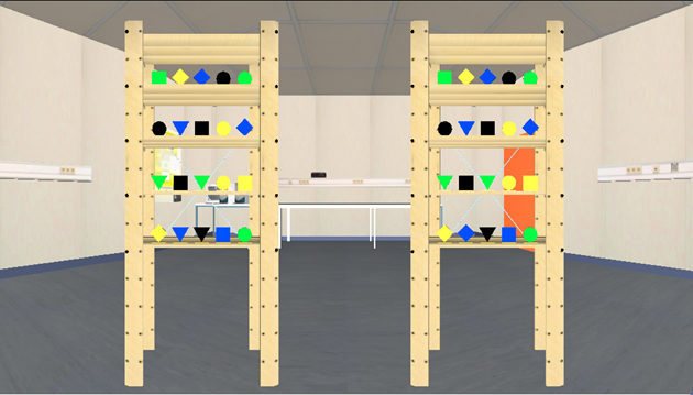
Fig.1: Screenshot of
the comparative visual search experiment. The subject
has to find the amount of differences in the object configuration
between the two cupboards as quickly and reliably as possible. The
amount of
differences in this example is 1 (first shelf from below; second
object from the left - blue triangle vs. blue diamond).
For this paradigm a virtual model of our VR-Lab was used as
environment. Thus, the experiment could be carried out later with real
cupboards and
objects in the real VR-room.
Movie 1: Illustration of the
real eye and head movements of a 'normal
subjects'
visual search.
Head movements are visualized by using a white rectangle. The red dot
represents the actual gaze
position. The width of the rectangle is +-25 degrees.
Eye movements remain almost within the range of the rectangle and are
not performed over the whole oculumotor range of +-55 degrees. The
subpression
of
the VOR (vestibulo-ocular reflex) is discernible during gaze shifts
between the both cupboards.
Movie 2:
Presentation of a 'hemianopic
patients' (complete visual
loss to the right) eye and head movements performend during an
experimental trial.
Head movements are visualized with the white rectangle and the
red dot
represents the actual gaze
position. The width of the rectangle is +-25 degrees.
This homonymous hemianopic patient compensates well (adequately) for the visual loss and performs only little head movements (small horizontal amplitude). As the only subject, this patient started with the upper right cupboard position for the comparative visual search. Interestingly, to the end of the trial the subject is changing the serach strategy from horizontal to vertical object scanning.
This homonymous hemianopic patient compensates well (adequately) for the visual loss and performs only little head movements (small horizontal amplitude). As the only subject, this patient started with the upper right cupboard position for the comparative visual search. Interestingly, to the end of the trial the subject is changing the serach strategy from horizontal to vertical object scanning.
Movie 3:
Presentation of a 'hemianopic
patients' (complete visual
loss to
the left) eye and head movements performend during an experimental
trial.
Head movements are visualized with the white rectangle and the
red dot
represents the actual gaze
position. The width of the rectangle is +-25 degrees.
This homonymous hemianopic patient shows an insufficent compensatory behavior. Whereas the head moves in a regular oscillatory manner between the left and the right cupboard, the eyes often move against the head and perform large and unprecise saccades. In contrast to the control subject (see above), this patients' head and eye movements are not harmonicly aligned.
This homonymous hemianopic patient shows an insufficent compensatory behavior. Whereas the head moves in a regular oscillatory manner between the left and the right cupboard, the eyes often move against the head and perform large and unprecise saccades. In contrast to the control subject (see above), this patients' head and eye movements are not harmonicly aligned.